")
")
Neologism recently introduced (2006) in dermatology coming from the fusion of two words: Entomology and Dermatoscopy.
The former points out the science that globally deals with the bugs, the latter concerns the discipline that analyzes the nevus and other cutaneous formations through a tool called dermatoscope that allows to observe the skin 10 times magnified.
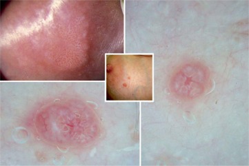
The Entodermoscopy (EDS) defines a new dermatoscopy application in the study (diagnosis and follow-up) of the cutaneous ectoparasitosis otherwise known as the human skin parasites diseases.
The infestations in which dermoscopy is more employed are the head and pubis lice, scabies, tungiasis, larva migrans and new others that are going to add.
The term officially was introduced in Bari on 02-03-2007 by Dr. Gaetano Scanni during a pediatric dermatologists progress on dermatoscopy , it have been taken back by Dr. Giuseppe Argenziano, an international dermatoscopy expert, in a publication titled Argenziano G. et All. Entodermoscopy: A New Tool for Diagnosing Skin Infections and Infestations; Dermatology 2008;216:14–23
In the text the author quotes the native source of word (Gaetano Scanni-Ernesto Bonifazi) and the relative work in which it was printed for the first time Scanni G. Bonifazi E. Viability of the head louse eggs in pediculosis capitis. A dermoscopy study ; Eur. J. Pediat. Dermatol. 2006; 16:201–204
The entodermoscopy (from others also said entomodermoscopy) is adding new informations on the complex interaction between the parasites and human skin. For example in the head lice field, it has allowed to underline previously unknown aspects about abortive nits and faces of this bug Scanni G. Bonifazi E. Feces of Pediculus capitis humanus as sign of viability of the louse; Eur. J. Ped. Dermatol. 17, 77-80, 2007
But it has also made easer the identification of endogenous keratinic structures called -horny cylinders- from real nits or to identify the exoskeleton, esuvia, that remains on the hair after three transformations that louse nymph overcome for becoming sexually mature adults Scanni G. Bonifazi E. Head Lice. Eur. J. Ped. Dermatol. Vol 18,33-64, 2008
The entodermoscopy advantage, unlike normal microscope, is to observe parasite in its own habitat showing aspects that in vitro would inevitably lost or strongly altered.
In this sense, the entodermoscopy proposes the spirit of the eco-ethological studies that describe animals and theirs environment interactions at the native state.
Currently, the entodermoscopy uses dermatoscopes that furnishes a standard 10x enlargement appropriate for the original applications it was set (control of the nevus and other cutaneous neoformations). On working about parasitosis it often required higher magnifications (200x or more), for this reason it is desirable to design new devices with more potentialities for the observer.
According to the Dr. Gaetano Scanni, lice specialist researcher, an ideal instrument for the cutaneous parasitosis study (entodermoscope) should possess at least the followings characteristics:
- Variability of enlargements from 10 to 200x through an optical zoom
- Photographic recording on a 5 megapixels or superior digital sensor
- Variable illumination through normal or polarized light
- Optional observation on immersion or dry mode without contact plaque
- Possiblity of inserting some micro tools into visual field
Dr. Gaetano Scanni
Dermatologist
2009-08-20