")
")
Entomology for dermatoscopists (entodermoscopy)
HUMAN SCABIES
Entodermoscopy.net , upload on 2012-02-06
Francesco Porcelli
Associate Professor, PhD, DiBCA Entomology and Zoology
Bari-Italy
Actually about fifteen Taxa of Acari (in their broad sense) are human ectoparasite either persistently associated or free-living, sometimes vectoring pathogenic microorganisms.
Just three species, namely Demodex folliculorum (Simon, 1842), D. brevis (Akbulatova, 1963) and Sarcoptes scabiei (L.), are common on man, being the formers found in hair follicle or sebaceous glands, while the latter living into skin. Moreover another mite, the Notoedres cati (Gerlach 1857), causes mange in cats and a transient dermatitis in man.
Scabies mite is a best-known mammal-noxious organism because it causes the mange in animals and the corresponding scabies in humans: a complex of itchy & scaly lesions consequent to its way of life. Scabies has been known for millennia, and has been referred to in the Old Testament of the Bible as well as by Aristotle. Scabies mites were described and drawn by the Italians Bonomo and Cestoni in 1689. This has been described as the first definitive description of a parasitic organism being responsible for an infectious disease and preceded modern germ theory by almost two centuries.
Definitive diagnosis of scabies requires microscopic visualization of a mite retrieved from a skin burrow.
S. scabiei is pleioxenous (1) and infests man and many others mammals species belonging to many different Genera. The single species S. scabiei is known to infest primates, horses, tapirs, wild and domestic bovid ruminants, pigs, camels, carnivores (including dogs and lions), rabbits and guinea pigs that are reservoirs for an eventual man infestation. Itch mite has the ability to pass among humans, among animals and from animals to man and vice-versa by close host-to-host contact or by fomites, i.e. bedding and clothing but just in case of crusted or Norwegian scabies.
Nevertheless due to a specific physiological parasite-host specialization, scabies can pass from horses or dogs (animal mange) to the humans but such infestations are mild and disappear spontaneously.Truly human scabies,sustained by Sarcoptes scabiei var. hominis, is widespread in the tropics but is not confined there.
Scabies mites can survive at least several days at moderate ambient temperatures in humid environments. Transmission via fomites as well as direct skin contact may therefore be occurring in nursing-home outbreaks and infections in hospital staff where a crusted scabies patient is the core transmitter.
Sarcoptes scabiei is a very small species, males and females being about 0.2 mm and 0.4 mm in length respectively, with striated cuticle and cutting chelicerae. The globose body bears various specialized dorsal scales and bristles. The body is opaque off-white in colour and the legs and mouthparts are brown. Adults and nymphs have eight legs and larvae have six.
The legs are arranged in two groups; the anterior pairs in both sexes end in stalked pulvilli, which, because of their disc-shaped appearance, are called suckers.
The pulvilli help the mite to grip the host's skin and so aid movement. In the female, the two posterior pairs of legs end in long bristles, but in the male only the third pair ends in bristles (the fourth resembling the first two pairs in having stalked pulvilli).
The lifecycle is simple: adult females deposit eggs as they precede in feeding tunnels, later eggs hatch thus releasing the larvae that moult in self bored short burrows and are found in hair follicles (2, 3). Nymphs are found in similar short “moulting pouches”. Males search for adult female in her moulting pouch where mating occurs. The entire life cycle takes 10-14 days.
The inseminated female scabies mite moves rapidly progressing up to 2.5 cm per minute, thus selecting a suitable site for burrowing. The burrow, usually about a centimetre long, is confined within the horny layer of the skin. Both sexes burrow but only the female makes permanent winding burrows, and the burrow of the male is no more than a millimetre long. Burrowing occurs at a rate of 0.5-5.0 mm per day
Most mite burrows occur in the interdigital and elbow skin, but skin of the scrotum, penis, breasts, knees and buttocks is also frequently infested; the face and scalp are rarely affected, except in children.Preferred sites for mites to burrow are where the stratum corneum is thin and soft and where there are few or no hair follicles. The more generalized skin rash,where there are no mites,is most commonly seen around the axillae, chest and abdomen, buttocks, and thighs.
A period of 3-4 weeks often elapses before itching, which is an immunologically mediated response to the presence of mites, mite faeces and dissolving skin enzymes, develops. Less than 20 mites are enough to produce intense itching (particularly at night). Relief by scratching can result in rupture of the mite burrows and removal or death of the mites.
While Sarcoptes scabiei infestation and mite reproduction in humans are usually self-limiting, a small minority develop hyperinfestation, with estimated total mite numbers in the most severe cases up to over a million. This debilitating condition is crusted or Norwegian scabies, so called because it was first described among leprosy patients in Norway in 1848. Crusted scabies has also been noted in overtly immunosuppressed patients, such as with malignancies, chemotherapy, and transplant patients. Most recently it has been increasingly recognized in advanced human immunodeficiency virus (HIV) infection. Crusted scabies also occasionally occurs in malnutrition, in Down’s syndrome, in the elderly and institutionalized, and in those with cognitive deficiency or physical debility who are unable to interpret properly, or respond to the itch by scratching.
The cardinal feature of crusted scabies is the development of hyperkeratotic skin crusts that may be loose, scaly, and flaky or thick and adherent. Skin flakes with thousands of mites can be shed daily on to bed linen or floors. While hands and feet are most commonly involved, the distribution is often extensive, including neck, scalp, and face as well as trunk and limbs, especially knees and elbows. Fissuring and secondary bacterial infection are common and regional lymphadenopathy is frequently present. A peripheral blood eosinophilia is common and serum levels of immunoglobulin E (IgE) are often extremely high (4). There is a high mortality in crusted scabies from secondary bacterial sepsis.
The most consistent factor associated with scabies is overcrowding, poverty plus poor hygiene andnutritional deficiencies are often associated with overcrowding, but the evidence that they are independent risk factors for scabies is mostly lacking.
Intrafamilial transmission as the commonest means of infection is supported by epidemiological studies and more recently by molecular studies showing the genotypes of mites from household members to be more homogenous than those from separate households within a community. Among adults sexual contact is an important means of transmission.
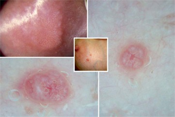
Scabies in adults and older children usually presents as an intensely pruritic rash, with the itch often worse at night. In primary infection the symptoms develop 3–4 weeks after infection. In those previously infected rash and itch can occur within 24–48 h of a new infection.In a minority of cases a nodular reaction (nodular scabies) occurs and it may persist for months after mite-eradicating treatment.
The classical diagnostic clinical sign of scabies is the burrow made by the adult female as it ingests and digests the horny layer of the epidermis. Burrows appear as serpiginous grayish, reddish, or brownish lines, 2–15 mm long. They are just visible to a trained unaided eye, but are difficult to see and classical burrows are often absent, even when the skin is inspected with magnification. Egg cases and mite fecal pellets are present inside the burrow. The papule at the burrow surface is usually small and erythematous, often excoriated or covered by a small blood clot. In infants and young children it can be vesicular or even bullous.
While in suspected scabies the presence of visible burrows or papules in the web spaces between the fingers is adequate for diagnosis, there is a wide range of clinical appearances like the inflammatory immune response, with any itchy generalized or local rash being possibly caused by scabies. Atypical appearances are more common in patients with longstanding scabies who may develop chronic excoriation and eczematization of limbs and trunk. Secondary bacterial infection of scabies lesions is frequent, with Streptococcus pyogenes and Staphylococcus aureus the common organisms. Scabies may therefore underly bacterial pyoderma, presenting as pustular or crusted sores, boils, cellulites, or lymphangitis/lymphadenitis.
Scabies should be considered in any patient presenting with a rash and itch. A recent history of scabies in contacts or household members should be sought and this increases the predictive value of a presumptive clinical diagnosis. Itch, which is worse at night, is also a useful predictor.
In suspected scabies the presence of visible burrows or papules/vesicles in the web spaces between the fingers is adequate for diagnosis. Magnifying glasses or dermatoscopes (5–10×) may help identify burrows. In any non-classical presentation it is recommended that skin samples be collected for microscopy if available.
Given the usually low number of mites present in scabies, the sensitivity of microscopy for confirming scabies is generally poor; diagnosis in unsuspected scabies will sometimes be made by histological examination of formalin-fixed skin biopsies of undiagnosed rashes.
Adult female mites reside in burrows within the stratum granulosum of the epidermis. Eggs, egg cases, mite fecal pellets, and debris are also present in the burrow. The associated underlying inflammatory response includes varying intensity and combinations of lymphocytes, histiocytes, eosinophils, and polymorphs. In crusted scabies there is massive expansion of the keratin layer due to proliferation keratinocytes. Numerous adult mites, eggs, and larvae are also usually seen, some clearly associated with burrows but many just spread throughout the epidermis and on the skin surface.
The mainstay of scabies therapy is topical acaricides, although oral ivermectin is increasingly being used in certain circumstances, most notably crusted scabies. Topical permethrin and oral ivermectin, where affordable, are currently considered the best treatment options.
Permethrin, a synthetic pyrethroid, is more expensive than other agents but is now considered the treatment of choice for scabies in many countries, including the UK and the USA. It has been recommended for use in children over 6 months, but is widely used in younger infants. However in vitro mite tolerance to permethrin has now been documented. The globally expanding resistance of head lice to permethrin suggests that continued surveillance for resistance in scabies is crucial.
Benzyl benzoate, an ester of benzoic acid and benzyl alcohol, has been an effective scabies treatment for more than 60 years. Although in vitro studies show that it kills scabies mites faster than permethrin, it has been traditionally recommended to leave the lotion on the skin for 24 h. The main problem with benzyl benzoate is skin irritation in the first minutes after application. Although the discomfort usually rapidly decreases in severity after several minutes, the burning sensation isnot uncommonly so painful that the lotion has to be washed off.
Crotamiton 10% cream has been widely used for scabies in children because of its low toxicity profile and the fact that it is well tolerated. However it has low and slow efficacy against S. scabiei and daily application for 3–5 days is recommended if it is to be used at all. It can be used for children less than 2 months of age if permethrin is not being used or is unavailable.
Sulphur compounds have been used to treat scabies for centuries. Although not active against scabies mites in vitro, when applied on the skin sulfur appears to elicit an active compound, possibly pentathionic acid, produced by skin microorganisms or epidermal cells. This may account for its apparent but limited efficacy. Sulphur creams are messy and smelly, can cause skin irritation, and require repeated application. Like crotamiton, they have a limited role, but 5% sulfur cream/lotion daily for 2–3 days is an alternative to permethrin in infants.
Although it is often recommended that topical therapy is only applied below the neck, scabies not infrequently involves the head and scalp in children and in the tropics also in adults, hence the general recommendation for whole-body application. The life cycle of S. scabiei supports repeat treatment at 1–2 weeks in more severe cases, to enable killing of nymphs hatched from eggs not killed by the initial treatment.
Ivermectin is a chemically modified avermectin with a half-life of 36h. It has been widely used for treatment of sarcoptic mange in animals in topical, oral, and parenteral preparations. Oral ivermectin for scabies is increasingly used worldwide, especially for crusted scabies. Although CNS toxicity from ivermectin is well documented in certain dog varieties, the extensive use in filarial programs has shown excellent tolerance, with few adverse reactions.
Concerns about toxicity and emerging resistance have resulted in a search for new topical scabicides. Promising agents include 5% tea tree oil, manufactured from the tree Melaleuca alternifolia.Tea tree oil is a traditional medicine used by indigenous Australians and has been shown to have excellent activity against a range of bacteria, including methicillin-resistant Staphylococcus aureus (MRSA), yeasts and herpes simplex virus. It rapidly kills Sarcoptes scabiei in vitro and has been successfully used with oral ivermectin in refractory crusted scabies.
Another essential oil recently successfully used for scabies is 20% lippia oil, extracted from the leaves of Lippia multiflora Moldenke, a shrub from the West African savannah. In both these essential oils terpenoids are the likely active components (4).
In small communities where scabies is endemic, whole-community treatment with topical permethrin has resulted in dramatic decreases in both scabies and streptococcal pyoderma. The success and sustainability of such programs require considerable planning, resources, and follow-up. Oral ivermectin rather than topical therapy has been used in those aged over 5 years in one successful program.
Cited bibliography
(1) Krantz, G.W. 2009 -Habits and Habitats. In “A manual of Acarology”, Krantz& Walter Eds.Texas Tech University Press, 807 pp.
(2) Varma, M.R.G. 1993 - Ticks and mites (Acari). In “Medical Insects and Arachnids”, Lane &Crosskey Eds.Chapman & Hall, 723 pp.
(3) Baker, E.W.; Evans, T.M.; Gould, D.J.; Hull, W.B.; Keegan, H.L. 1956 - A Manual of Parasitic Mites of Medical or Economic Importance. A Technical Publication of the National Pest Control Association, Inc. New York, 170 pp.
(4) Currie, B.; Hengge, U.R. 2006 - Scabies. In: “Tropical Dermatology” Tyring, Lupi&Hengge Eds. Elsevier Churchill Livingstone, 515 pp.
Selected bibliography
Baker, A.S. 1999 - Mites and ticks of domesticanimals an Identification Guide and Information Source. The Natural HistoryMuseum, The Stationery Office, 240 pp.
Barriga, O.O. 2003 - Zoonoses and Communicable Diseases Common to Man and Animals IIIParasitoses, 3rd Ed. Pan American Health Organization, 395 pp.
Buxton, P.K.; Morris-Jones, R. 2009 - ABC of Dermatology, 5th ed. Wiley-Blackwell, 210 pp.
Guerrant, R.L.; Walker, D.H.; Weller, P.F. 2006 - Tropical Infectious Diseases Principles, Pathogens and Practice, 3rd Ed., Saunders Elsevier, 1023 pp.
Hunter, J.A.A.; Savin, J.A.; Dahl, M.V. 2002 - ClinicalDermatology, 3rd Ed. BlackwellScience, 365 pp.
James, W.D.; Berger, T.G.; Elston, D.M. 2011 - Andrews’ Diseases of the SkinClinicalDermatology, 11th Ed. SaudersElsevier, 959 pp.
Lapage, G. 1963 -AnimalParasitic in Man. Dover Publications, 320 pp.
Meinking, T.L.; Burkhart, C.N.; Burkhart, C.G.; Elgart, G.2008 – Infestations. In “Dermatology 2nd Ed.” Bolognia, Jorizzo&RapiniEds.MosbyElsevier, 1319 pp.
Oconnor, B.M. 2009 - Cohort Astigmatina. In “A manual of Acarology”, Krantz& Walter Eds.Texas Tech University Press, 807 pp.
Patton, W.S.; Francis, W.C. 1913 - A Textbook of MedicalEntomology. Christian Literature Society for India, 764 pp.
Weese, J.S.; Fulford, M.B. 2011 - Companion AnimalZoonoses. Blackwell Publishing, 319 pp.